
Brain Tumor Classification from MRI Images Using Pretrained Deep Convolutional Neural Networks
Keywords:
Brain tumor, convolutional neural networks, detection and localization, MRI scans, pre-trained model.Abstract
Brain tumor is an abnormal tissue growth which may lead to cancer and it is characterized by the excessive cell proliferation in certain parts of the brain. One of the current reliable technologies that may be employed to identify brain tumor is to apply Magnetic Resonance Imaging (MRI) scans. The scanned MRI images are then monitored and conventionally examined by the medical specialists to observe the existence of tumor. As the number of people suffering brain tumor is very much increased and their corresponding mortality rate has reached 18,600 in the year 2021, research on devising more effective and efficient tools to assist the medical specialists on the identification of brain tumor is considered very urgent. In the previous studies, the Convolutional Neural Network (CNN) based models demonstrate their capability to detect brain tumor with 96% classification accuracy and they are found to be more reliable than other machine learning based models. In an attempt to obtain the best classification accuracy on both binary and multi-class MRI brain images, some powerful pretrained deep CNN models namely VGG16, VGG19, ResNet50, ResNet101, and InceptionResNetV2 are computationally experimented using publicly open MRI datasets. State of the art accuracy of the pretrained models is achieved by fine tuning the parameters of the convolutional layers of the base models and followed by feeding the high-level feature maps extracted from each corresponding base model either into flatten layers or into global average pooling layers prior to classifying the tumor by fully connected layers. The highest testing accuracy score as high as 99% is achieved by the VGG16 and InceptionResnetV2 on binary MRI image classification and a little higher than 99% is obtained by all the pretrained models on multi-class MRI brain image classification.
Downloads
References
SEER, “Surveillance, Epidemiology, and End Results Program.”
S. A. H. S. Javadi and B. Rezaei, “Brain tumors and indications for brain imaging in patients with psychiatric manifestations: a case report,” Middle East Curr. Psychiatry, vol. 28, no. 1, pp. 0–4, 2021, doi: 10.1186/s43045-021-00136-2.
A. Çinar and M. Yildirim, “Detection of tumors on brain MRI images using the hybrid convolutional neural network architecture,” Med. Hypotheses, vol. 139, no. February, p. 109684, 2020, doi: 10.1016/j.mehy.2020.109684.
H. Mzoughi et al., “Deep Multi-Scale 3D Convolutional Neural Network (CNN) for MRI Gliomas Brain Tumor Classification,” J. Digit. Imaging, vol. 33, no. 4, pp. 903–915, 2020, doi: 10.1007/s10278-020-00347-9.
Y. Bhanothu, A. Kamalakannan, and G. Rajamanickam, “Detection and Classification of Brain Tumor in MRI Images using Deep Convolutional Network,” 2020 6th Int. Conf. Adv. Comput. Commun. Syst. ICACCS 2020, pp. 248–252, 2020, doi: 10.1109/ICACCS48705.2020.9074375.
R. L. Kumar, J. Kakarla, B. V. Isunuri, and M. Singh, “Multi-class brain tumor classification using residual network and global average pooling,” Multimed. Tools Appl., vol. 80, no. 9, pp. 13429–13438, 2021, doi: 10.1007/s11042-020-10335-4.
K. N. Qodri, I. Soesanti, and H. A. Nugroho, “Image Analysis for MRI-Based Brain Tumor Classification Using Deep Learning,” IJITEE (International J. Inf. Technol. Electr. Eng., vol. 5, no. 1, p. 21, 2021, doi: 10.22146/ijitee.62663.
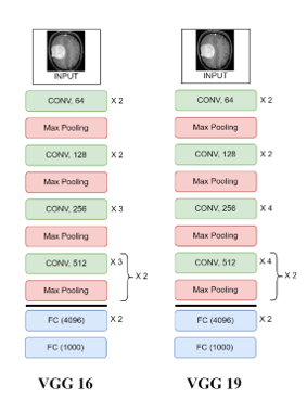
K. Simonyan and A. Zisserman, “Very deep convolutional networks for large-scale image recognition,” 3rd Int. Conf. Learn. Represent. ICLR 2015 - Conf. Track Proc., pp. 1–14, 2015.
K. He, X. Zhang, S. Ren, and J. Sun, “Deep residual learning for image recognition,” Proc. IEEE Comput. Soc. Conf. Comput. Vis. Pattern Recognit., vol. 2016-Decem, pp. 770–778, 2016, doi: 10.1109/CVPR.2016.90.
C. Szegedy, S. Ioffe, V. Vanhoucke, and A. A. Alemi, “Inception-v4, inception-ResNet and the impact of residual connections on learning,” 31st AAAI Conf. Artif. Intell. AAAI 2017, vol. 42, no. 1, pp. 4278–4284, 2017.
Downloads
Published
How to Cite
Issue
Section
License

This work is licensed under a Creative Commons Attribution-ShareAlike 4.0 International License.
All papers should be submitted electronically. All submitted manuscripts must be original work that is not under submission at another journal or under consideration for publication in another form, such as a monograph or chapter of a book. Authors of submitted papers are obligated not to submit their paper for publication elsewhere until an editorial decision is rendered on their submission. Further, authors of accepted papers are prohibited from publishing the results in other publications that appear before the paper is published in the Journal unless they receive approval for doing so from the Editor-In-Chief.
IJISAE open access articles are licensed under a Creative Commons Attribution-ShareAlike 4.0 International License. This license lets the audience to give appropriate credit, provide a link to the license, and indicate if changes were made and if they remix, transform, or build upon the material, they must distribute contributions under the same license as the original.


