Developing a Methodology for the Formation of a System of Attributes of Pathological Vascular Changes in the Fundus
Keywords:
classification methods, clustering methods, discriminant analysis, evidence-based medicine, vascular pathology diagnosisAbstract
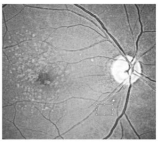
This research aimed to develop a methodology for extracting data from diagnostic images of the fundus blood vessels and methods for their high-precision evaluation, focused on ensuring the diagnostic process standardization, reducing the time of examination and its cost within the framework of evidence-based medicine. Timely and competent diagnosis plays an important role in obtaining an optimal result for treating vascular pathologies. This research evaluated the effectiveness of existing approaches to the analysis of the geometric diagnostic attributes of the fundus vascular system state reflected in the images, which are necessary for identifying pathological vascular changes. A technique was developed for the formation of an optimal system of geometric diagnostic attributes according to the criterion of separability. It was shown that the most effective method for solving this problem is the discriminant analysis method, which, in the presence of a strong connection between certain groups of attributes, makes it possible to decide whether it is expedient to use them and reduce the dimension of the attribute space. Reducing the dimension can significantly reduce the number of calculations. The results of using full-scale images of the fundus with active medical support confirmed the effectiveness of the developed technique.
Downloads
References
S. Biswas and Y. Hasija, “Chapter 6 - Big data analytics in precision medicine”, In Big Data Analytics for Healthcare, P. Keikhosrokiani, Ed., New-York, USA: Academic Press, 2022.
V. A. Gorelov, E. Y. Linskaya, A. A. Tatarkanov, I. A. Alexandrov, and S. A. Sheptunov, “Complex Methodological Approach to Introduction of Modern Telemedicine Technologies into the Healthcare System on Federal, Regional and Municipal Levels”, presented at the 2020 International Conference Quality Management, Transport and Information Security, Information Technologies, Yaroslavl, Russia, 2020.
M. Chaochao, W. Xinlu, W. Jie, C. Xinqi, X. Liangyu, X. Fang, and Q. Ling, “Real-world big-data studies in laboratory medicine: Current status, application, and future considerations”, Clinical Biochemistry, vol. 84, pp. 21-30, 2020.
S. Khodaei, R. Sadeghi, P. Blanke, J. Leipsic, A. Emadi, and Z. Keshavarz-Motamed, “Towards a non-invasive computational diagnostic framework for personalized cardiology of transcatheter aortic valve replacement in interactions with complex valvular, ventricular and vascular disease”, International Journal of Mechanical Sciences, vol. 202–203, art. 106506, 2021.
A. D. Choi, H. Marques, V. Kumar, W. F. Griffin, H. Rahban, R. P. Karlsberg, R. K., Zeman, R. J. Katz, and J. P. Earls, “CT Evaluation by Artificial Intelligence for Atherosclerosis, Stenosis and Vascular Morphology (CLARIFY): A Multi-center, international study”, Journal of Cardiovascular Computed Tomography, vol. 15, no. 6, pp. 470-476, 2021.
T. Montalcini, et al., “Brachial artery diameter measurement: A tool to simplify non-invasive vascular assessment”, Nutrition, Metabolism and Cardiovascular Diseases, vol. 22, no. 1, pp. 8-13, 2012.
P. Guimarães, et al., “Ocular fundus reference images from optical coherence tomography”, Computerized Medical Imaging and Graphics, vol. 38, no. 5, pp. 381-389, 2014.
V. Romano et al., “Imaging of vascular abnormalities in ocular surface disease”, Survey of Ophthalmology, vol. 67, no. 1, pp. 31-51, 2022.
M. Kouroupis, N. Korfiatis, and J. Cornford, “Chapter 11 - Artificial intelligence–assisted detection of diabetic retinopathy on digital fundus images: concepts and applications in the National Health Service”, in “Innovation in Health Informatics”, A. Lytras, Ed., New-York, USA: Academic Press, 2020.
M. A. U. Khan et al., “Thin Vessel Detection and Thick Vessel Edge Enhancement to Boost Performance of Retinal Vessel Extraction Methods”, Procedia Computer Science, vol. 163, pp. 618-638, 2019.
S. Thainimit, et al., “Robotic process automation support in telemedicine: Glaucoma screening usage case”, Informatics in Medicine Unlocked, vol. 31, art. 101001, 2022.
A. Naranjo, et al., “Retinal artery and vein occlusion in calciphylaxis”, American Journal of Ophthalmology Case Reports, vol. 26, art. 101433, 2022.
G. M. Trovato, “Eyeing the retinal vessels: A window on the heart and beyond”, Atherosclerosis, vol. 348, pp. 51-52, 2022.
W. Magoń, et al., “Right Ventricular Epicardial Vascularisation in Patients with Pulmonary Arterial Hypertension”, Heart, Lung and Circulation, vol. 27, no. 12, pp. 1428-1436, 2018.
M. Jeub, et al., “Sonographic assessment of the optic nerve and the central retinal artery in idiopathic intracranial hypertension”, Journal of Clinical Neuroscience, vol. 72, pp. 292-297, 2020.
R. Simo and S. Frontoni, “Neuropathic damage in the diabetic eye: clinical implications”, Current Opinion in Pharmacology, vol. 55, pp. 1-7, 2020.
A. Chehaitly, et al., “Flow-mediated outward arterial remodeling in aging”, Mechanisms of Ageing and Development, vol. 194, art. 111416, 2021.
S. Kihira, et al., “Trans-synaptic degeneration of the optic radiation from optic nerve atrophy”, Radiology Case Reports, vol. 16, no. 4, pp. 855-857, 2021.
G. Uludag, A. Onay, and S. Onal, “Unilateral paraneoplastic optic disc edema and retinal periphlebitis in pineal germinoma”, American Journal of Ophthalmology Case Reports, vol. 10, pp. 236-239, 2018.
H. Jamoussi, et al., “Relapse of acute lymphoblastic leukemia revealed by an optic neuropathy”, Revue Neurologique, vol. 176, 131-136, 2020.
J. Han, Y. Wang, and H. Gong, “Fundus Retinal Vessels Image Segmentation Method Based on Improved U-Net”. IRBM, in press, 2022.
B. J. Palermo, et al., “Sensitivity and specificity of handheld fundus cameras for eye disease: A systematic review and pooled analysis”, Survey of Ophthalmology, vol. 67, no. 5, pp. 1531-1539, 2021.
M. Monjur et al., “Smartphone based fundus camera for the diagnosis of retinal diseases”, Smart Health, vol. 19, art. 100177, 2021.
A. Tatarkanov, I. Alexandrov, and R. Glashev, “Synthesis of Neural Network Structure for the Analysis of Complex Structured Ocular Fundus Images”, Journal of Applied Engineering Science, vol. 19, no. 2, pp. 344-355, 2021.
A. Tatarkanov, I. Alexandrov, and I. Vorobieva, “Automated Diagnostics of Early Stages of Diabetic Retinopathy using Neural Network Modeling Methods”, in Proc. 2021 International Conference on Quality Management, Transport and Information Security, Information Technologies, Yaroslavl, Russia, 06-10 September 2021.
A. Quarteroni, A. Veneziani, and C. Vergara, “Geometric multiscale modeling of the cardiovascular system, between theory and practice”, Computer Methods in Applied Mechanics and Engineering, vol. 302, pp. 193-252, 2016.
A. Skiadopoulos, P. Neofytou, and C. Housiadas, “Comparison of blood rheological models in patient specific cardiovascular system simulations”, Journal of Hydrodynamics, vol. 29, no. 2, pp. 293-304, 2016.
X. Liu, et al., “Quantitative assessment of retinal vessel density and thickness changes in internal carotid artery stenosis patients using optical coherence tomography angiography”, Photodiagnosis and Photodynamic Therapy, vol. 39, art. 103006, 2022.
P. F. Sharp et al., “The Value of Digital Imaging in Diabetic Retinopathy”, Health Technology Assessment, vol. 7, no. 30, 2003.
T. Salahuddin and U. Qidwai, “Computational methods for automated analysis of corneal nerve images: Lessons learned from retinal fundus image analysis”, Computers in Biology and Medicine, vol. 119, art. 103666, 2020.
M. M. Fraz, et al., “Blood vessel segmentation methodologies in retinal images”, Computer Methods and Programs in Biomedicine, vol. 108, no. 1, pp. 407-433, 2012.
Y. Wang, et al., “Retinal vessel segmentation using multiwavelet kernels and multiscale hierarchical decomposition”, Pattern Recognition, vol. 46, no. 8, pp. 2117-2133, 2013.
T. Li, et al., “Applications of deep learning in fundus images: A review”, Medical Image Analysis, vol. 69, art. 101971, 2021.
R. D. Párizs, et al. “Machine Learning in Injection Molding: An Industry 4.0 Method of Quality Prediction”, Sensors, vol. 22, no. 7, art. 2704, 2022.

Downloads
Published
How to Cite
Issue
Section
License
Copyright (c) 2023 Aslan Adal’bievich Tatarkanov, Abas Khasanovich Lampezhev, Ruslan Khalitovich Tekeev, Dmitry Alekseevich Marenkov, Leonid Mikhailovich Chervyakov

This work is licensed under a Creative Commons Attribution-ShareAlike 4.0 International License.
All papers should be submitted electronically. All submitted manuscripts must be original work that is not under submission at another journal or under consideration for publication in another form, such as a monograph or chapter of a book. Authors of submitted papers are obligated not to submit their paper for publication elsewhere until an editorial decision is rendered on their submission. Further, authors of accepted papers are prohibited from publishing the results in other publications that appear before the paper is published in the Journal unless they receive approval for doing so from the Editor-In-Chief.
IJISAE open access articles are licensed under a Creative Commons Attribution-ShareAlike 4.0 International License. This license lets the audience to give appropriate credit, provide a link to the license, and indicate if changes were made and if they remix, transform, or build upon the material, they must distribute contributions under the same license as the original.


