
Optimization of Image Analysis Algorithm for the Diagnosis of Brain Tumors
Keywords:
Artificial Neural Network (ANN), Carcinoma, Diagnostics, MR Images, Particle Swarm Optimization (PSO), Segmentation, TumorAbstract
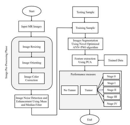
Image analysis is the best diagnostics tool for carcinoma cell diagnosis. Symptomatic clinical correlation with pathological findings amalgamated with Image analysis is the best way to diagnose cancerous cells and their nature. The worldwide brain tumor recorded cases have risen in recent years which is a huge challenge for clinicians to detect the tumor in the early stage of development. Though, plenty of research has been conducted on brain MR image segmentation and features extraction, but the existing approaches comprise certain limitations. These approaches are not restricted to massive time consumption, the lower rate of accuracy along with the more computational cost is also challenging for current scenario. In this research, a novel and optimized image analysis algorithm for the diagnosis of brain tumors has been presented. This optimized image analysis algorithm is rooted in the combined artificial neural network (ANN) as well as particle swarm optimization (PSO) approach. The obtained results have been recorded enhanced in terms of accuracy as well as sensitivity in comparison to the existing brain tumor detection methods. Using the combined approach i.e., ANN and PSO methods, the accuracy, sensitivity as well as recorded time was optimal. The proposed optimized image analysis algorithm takes very less time in operation i.e., only 0.8 seconds which is minimal in comparison with earlier methods. The measured accuracy and sensitivity for the proposed optimized image analysis algorithm on white matter (WM), gray matter (GM), and tumor MR images are found 98%, 97%, and 99% as well as 97.5%, 98.4%, and 99.22%, respectively.
Downloads
References
V. Ntziachristos, M. A. Pleitez, S. Aime, and K. M. Brindle, “Emerging Technologies to Image Tissue Metabolism,” Cell Metabolism, vol. 29, no. 3, pp. 518-538, 2019. doi: 10.1016/j.cmet.2018.09.004.
S. Gudise, G. B. Kande, and T. S. Savithri, “MR Brain Image Segmentation to Detect White Matter, Gray Matter, and Cerebro Spinal Fluid Using TLBO Algorithm,” Int. J. Image Graph., vol. 21, no. 3, 2021, doi: 10.1142/S0219467821500340.
S. Pontes-Filho, A. G. Dahl, S. Nichele, and G. B. M. e. Mello, “A Deep Learning-Based Tool for Automatic Brain Extraction from Functional Magnetic Resonance Images of Rodents,” in Lecture Notes in Networks and Systems, 2022. doi: 10.1007/978-3-030-82199-9_36.
M. K. Abd-Ellah, A. I. Awad, A. A. M. Khalaf, and H. F. A. Hamed, “A review on brain tumor diagnosis from MRI images: Practical implications, key achievements, and lessons learned,” Magnetic Resonance Imaging., vol. 61, pp. 300-318, 2019. doi: 10.1016/j.mri.2019.05.028.
C. E. Zimmerman et al., “Automatic Segmentation of Bone Selective MR Images for Visualization and Craniometry of the Cranial Vault,” Acad. Radiol., 2021, doi: 10.1016/j.acra.2021.03.010.
H. Sajedi and N. Pardakhti, “Age Prediction Based on Brain MRI Image: A Survey,” Journal of Medical Systems. 2019. doi: 10.1007/s10916-019-1401-7.
I. Oksuz, “Brain MRI artefact detection and correction using convolutional neural networks,” Comput. Methods Programs Biomed., 2021, doi: 10.1016/j.cmpb.2020.105909.
Z. Zhang, J. Li, C. Tian, Z. Zhong, Z. Jiao, and X. Gao, “Quality-driven deep active learning method for 3D brain MRI segmentation,” Neurocomputing, vol. 446, pp. 106-117, 2021, doi: 10.1016/j.neucom.2021.03.050.
Z. Jiang, X. Lyu, J. Zhang, Q. Zhang, and X. Wei, “Review of deep learning methods for MRI brain tumor image segmentation,” Journal of Image and Graphics. 2020. doi: 10.11834/jig.190173.
I. Abd El Kader et al., “Brain Tumor Detection and Classification on MR Images by a Deep Wavelet Auto-Encoder Model,” Diagnostics, vol. 11, no. 9, 2021, doi: 10.3390/diagnostics11091589.
A. Fawzi, A. Achuthan, and B. Belaton, “Brain image segmentation in recent years: A narrative review,” Brain Sciences. 2021. doi: 10.3390/brainsci11081055.
Y. Zhang, S. Wang, G. Ji, and Z. Dong, “An MR brain images classifier system via particle swarm optimization and kernel support vector machine,” Sci. World J., 2013, doi: 10.1155/2013/130134.
M. Ahmadi, A. Sharifi, M. Jafarian Fard, and N. Soleimani, “Detection of brain lesion location in MRI images using convolutional neural network and robust PCA,” Int. J. Neurosci., 2021, doi: 10.1080/00207454.2021.1883602.
D. Filatov and G. N. A. H. Yar, “Brain Tumor Diagnosis and Classification via Pre-Trained Convolutional Neural Networks,” 2022, [Online]. Available: http://arxiv.org/abs/2208.00768
V. Sheejakumari and B. Sankara Gomathi, “MRI brain images healthy and pathological tissues classification with the aid of improved particle swarm optimization and neural network,” Comput. Math. Methods Med., 2015, doi: 10.1155/2015/807826.
M. M. Badža and M. C. Barjaktarović, “Classification of brain tumors from mri images using a convolutional neural network,” Appl. Sci., vol. 10, no. 6, 2020, doi: 10.3390/app10061999.
A. K. Sharma, A. Nandal, A. Dhaka, D. Koundal, D. C. Bogatinoska, and H. Alyami, “Enhanced Watershed Segmentation Algorithm-Based Modified ResNet50 Model for Brain Tumor Detection,” Biomed Res. Int., 2022, doi: 10.1155/2022/7348344.
V. Sivakumar and N. Janakiraman, “A novel method for segmenting brain tumor using modified watershed algorithm in MRI image with FPGA,” BioSystems, vol. 198, 2020, doi: 10.1016/j.biosystems.2020.104226.
I. Aboussaleh, J. Riffi, A. M. Mahraz, and H. Tairi, “Brain tumor segmentation based on deep learning’s feature representation,” J. Imaging, vol. 7, no. 12, 2021, doi: 10.3390/jimaging7120269.
R. Ranjbarzadeh, A. Bagherian Kasgari, S. Jafarzadeh Ghoushchi, S. Anari, M. Naseri, and M. Bendechache, “Brain tumor segmentation based on deep learning and an attention mechanism using MRI multi-modalities brain images,” Sci. Rep., 2021, doi: 10.1038/s41598-021-90428-8.
Downloads
Published
How to Cite
Issue
Section
License

This work is licensed under a Creative Commons Attribution-ShareAlike 4.0 International License.
All papers should be submitted electronically. All submitted manuscripts must be original work that is not under submission at another journal or under consideration for publication in another form, such as a monograph or chapter of a book. Authors of submitted papers are obligated not to submit their paper for publication elsewhere until an editorial decision is rendered on their submission. Further, authors of accepted papers are prohibited from publishing the results in other publications that appear before the paper is published in the Journal unless they receive approval for doing so from the Editor-In-Chief.
IJISAE open access articles are licensed under a Creative Commons Attribution-ShareAlike 4.0 International License. This license lets the audience to give appropriate credit, provide a link to the license, and indicate if changes were made and if they remix, transform, or build upon the material, they must distribute contributions under the same license as the original.


